What are some diagnostic tests for nervous system disorders?
Looking for damage to the nervous system is complex. Many of the same symptoms can happen in different disorders. Many disorders also don't have clear-cut causes or tests. That can make a diagnosis even harder.
To diagnose a nervous system disorder, a doctor starts with a complete medical history and physical exam. They may also use one or more of these tests:
- CT scan. This test uses X-rays and a computer. Images are created of any part of the body. CT scans are more detailed than general X-rays. They are used to find problems of the brain, spine, or other parts of the nervous system.
- Electroencephalogram (EEG). This test records the brain's activity through electrodes placed on the scalp.
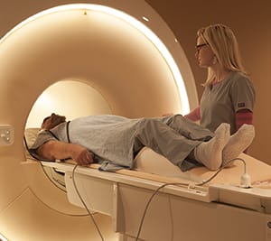
- MRI. Large magnets, radio waves, and a computer make images of organs and structures. MRI offers much more detail than CT scan, without radiation.
- Electrodiagnostic tests. These include electromyography (EMG) and nerve conduction velocity (NCV). These tests are used to find problems of the muscles and motor neurons. Electrodes are placed into the muscle or on the skin over a muscle or muscle group. Electrical activity and muscle response are recorded.
- Positron emission tomography (PET). This test uses a small amount of radioactive material, a camera, and a computer. It can show how well organs and tissues are working. It may find the early onset of disease before imaging tests can.
- Arteriogram (angiogram). This is an X-ray of the arteries and veins. It can show blockage or narrowing of the blood vessels.
- Spinal tap (lumbar puncture). A special needle is placed into the lower back, into the spinal canal. This is the area around the spinal cord and nerves. The pressure in the spinal canal and brain is measured. A small amount of cerebrospinal fluid (CSF) can be removed. The sample is sent for testing to find out if there is an infection or other problems. CSF is the fluid that bathes the brain and spinal cord.
- Evoked potentials. This test records the brain's electrical response to visual, hearing, and sensory stimuli.
- Myelogram. Dye is injected into the spinal canal to make it visible on X-rays. This test is used less commonly because MRI is widely available.
- Neurosonography. This uses ultra-high-frequency sound waves. It allows the doctor to study blood flow in cases of possible stroke. This includes carotid ultrasound and transcranial doppler.
- Ultrasound (sonography). This test uses high-frequency sound waves and a computer to make images of blood vessels, tissues, and organs. Ultrasounds are used to view internal organs as they work. They also assess blood flow through vessels.
- Biopsy. In this procedure, a small amount of tissue is removed. It's usually done during brain surgery or on its own to learn more about a tumor.
© 2000-2026 The StayWell Company, LLC. All rights reserved. This information is not intended as a substitute for professional medical care. Always follow your healthcare professional's instructions.